

MedSurgPI, LLC partners with Exquisite Biomedical Consulting to provide Medical Affairs and Medical Communication services to the Canadian pharmaceutical, biotechnology, medical device and related technology companies and institutions.
Exquisite Biomedical Consulting, based in Vancouver, British Columbia offers a comprehensive suite of Medical Affairs and Medical Communication Services such as Medical Affairs Strategy, KOL and publication strategy, advisory panel strategy and execution, MSL training, due diligence for business development, medical writing of Clinical Evaluation Reports (EU MDR and MDD), white papers and manuscripts.
MedSurgPI offers part time Chief Medical Officer, Medical Monitoring, and Medical Consulting for Product Development and Medical Affairs for Drug, Biologic, and Device companies.
In addition, Shabnam Vaezzadeh, MD, MPA, BCMAS, CEO of Exquisite Biomedical Consulting will head up the medical writing and publications groups for MedSurgPI.
For more information contact Exquisite Biomedical Consulting: https://lnkd.in/dYV4zGbC / contact@exquisitebiomedical.com. MedSurgPI info: www.medsurgpi.com / svanvactor@medsurgpi.com.
The Case for Digital Pill Use in Clinical Trials
Clinical Trials and Practice - Open Tournal (CTPOJ)
by Gerald L. Klein, MD / Published August 17, 2021

Abstract: Medication adherence in clinical trials is significantly overestimated through every phase of drug development. This can cause a reduction in statistical power, potentially resulting in incorrect conclusions regarding efficacy, safety, tolerability, and dose-response relationships, in addition to major cost overruns. Digital pill systems enable adherence measurement through an embedded ingestible sensor paired with an external receiver. An oral pharmaceutical product is over-encapsulated by a pharmaceutical-grade shell containing a biocompatible sensor. Upon exposure to gastrointestinal fluid, the shell dissolves and the sensor is activated. Medication ingestion data is transmitted via a digital signal. Clinicians and researchers use this data to track, in real time, when and if a medication was taken. These systems have demonstrated a 99.4% rate of accuracy, and have over 15-years of supporting experience and safety data. Spurred by the accelerated adoption of technology in healthcare and in everyday life, patients have become tech-savvy. They quickly adapt to these devices, and are able to use them safely and effectively. Digital pills can be implemented in most types of studies. In early-stage trials such as pharmacokinetic and pharmacodynamic studies, or dose-finding studies, accurate information on maximum-tolerated dose levels is essential and cannot be established unless study participants are highly adherent. In later-stage pivotal trials, effective medication adherence tracking can strengthen the dataset and confidence in the study results. Significant nonadherence may generate results that do not meet statistical or clinical significance for the critical endpoints, resulting in at worst, a failed trial, or at best, the need to enroll additional patients at substantial additional cost. Most clinical trials fail to achieve statistical significance, and poor medication adherence is often an important contributor. A digital pill system can ensure the quality and integrity of adherence data, increase confidence in the overall study data, and improve clinical trial efficiency.
The Importance of Teaching and Fostering Clinical Research in Primary Health care
Gerald L. Klein, MD and Mark A. Brown, PhD
It is important for primary health care (PHC) teaching institutions and hospitals to create an atmosphere fostering clinical investigation for all health care practitioners (HCP). This involves not just clinical trials, but observations, examinations, and investigations are a critical part of the education of any health science students. This is the basis of science. Without knowing how to apply a scientific thought process and methodology to a clinical situation, it will prevent one from reaching their optimum abilities. Continue reading…

White Paper Published in DIA

The Extraordinary Problem of Medication Nonadherence in Oncology Patients and How Digital Pills Can Make a Difference. Available to download.
Recognizing High Risk Traumatic Wounds and Preventing Infections and Complications
Review Article / Emergency Medicine and Trauma Care Journal
by Gerry L. Klein and Peter C. Johnson, MedSurgPI, LLC

Abstract: Simple traumatic wounds are a frequent event that can usually be managed without sequelae, unless the wound is of high risk. High risk wounds have a greater propensity to become infected and complicated. Such wounds are characterized by a specific type of wound (i.e. jagged), location of the wound (i.e. lower leg); and patient’s underlying medical condition (i.e: diabetes).If these wounds become infected, they have a negative impact on morbidity, mortality, quality of life, and costs. The take-away should be a wake-up call to physicians specifically and healthcare professionals more broadly that a much more aggressive and effective treatment regimen to prevent wounds from becoming infected is required. Such a regimen should likely include a comprehensive understanding of wound types, the degrees of microbial contamination, and novel ways to prevent infections through wound debridement and irrigation.
Dr. Gerry Klein to serve as panelist on Webinar: Wound Infection in Humanitarian Emergencies

#PrepareToProtect #IRPWebinar #MedicalResponse #HumanitarianEmergencies #GFARC #WHO
We at MedSurgPI, LLC are delighted to participate in and invite you to a webinar on wound infection, organized by Infection Reduction Partners, an initiative to promote and champion cross-sector collaboration and partnerships towards the effective reduction of infection rates worldwide.
This webinar will explore the cross-sector data, guidelines, and best practices in wound infection during disaster response and humanitarian emergencies. A presentation of practical and innovative options to reduce the risk of infection will be shared and we will discuss current publications including IFRC’s “International First Aid, Resuscitation and Education Guidelines 2020,” WHO’s Guidance, “Prevention and Management of Wound Infection,” WHO/ICRC’s “Management of Limb Injuries,” and WHO’s “Global Guidelines for the Prevention of Surgical Site Infection”.
Panelists:
- Dr. Pascal Cassan, MD, Head of Global First Aid Reference Centre, International Federation of Red Cross and Red Crescent Societies
- Flavio Salio, Team Lead - Emergency Medical Teams at the World Health Organization
- Kim Delahanty, BSN, PHN, MBA/HCM, CIC, FAPIC, Infection Prevention Control Advisor/Referent, Médecins Sans Frontieres USA/OCP (Doctors without borders)
- Dr. Gerald L. Klein, MD, Principal at MedSurgPI, LLC, wound infection consultant
Moderator:
Tessy Antony de Nassau, Ambassador for UNAIDS, and Patron to UNA-UK
Find out more on IRP social media Facebook, YouTube, Twitter, Instagram, LinkedIn!
Audience:
Medical team members involved in humanitarian aid, disaster relief, and emergency response: Medical directors, doctors, nurses, technicians, responders, infection preventionists, health officers, medical team leaders
NGOs
International Organizations
Government/Ministries of Health
Potential Psychological Benefits of a Regenerative Graft for Nipple Reconstruction

Link to this article appearing in the Journal of Aesthetic & Reconstructive Surgery ISSN 2472-1905
by Gerald L. Klein and Peter C. Johnson
Vol. 7 No.2:17
The Medical Monitor is one of the key players in keeping subjects safe in clinical trials. This article helps explore the role of the Medical Monitor
Journal of Clinical Research Best Practices
Medical Monitoring of Clinical Research Studies
By Gerald L. Klein, Peter C. Johnson, and Roger Morgan

Introduction
The Medical monitor’s (MM’s) primary responsibilities in a clinical trial are to oversee the safety and protection of the research subjects and to provide independent oversight to help ensure the scientific reliability, clinical integrity, and quality of the clinical trial. Although the Food and Drug Administration (FDA) has not spelled out the necessity and role of the medical monitor, MM participation is important for compliance with Good Clinical Practice (GCP) guidelines and MMs almost always play a significant role in multicenter clinical trials.1 The best practices for MMs described in this article can also help study sponsors comply with International Council for Harmonization (ICH) guidelines, Technical Requirements for Pharmaceuticals for Human Use, the United States Code of Federal Regulations (CFR), and FDA regulations and guidances.2
The MM collaborates with the project manager, safety, data management, biostatistics and quality departments, principal and sub-investigators, the site coordinator, the Data & Safety Monitoring Board (DSMB), and any other group that directly or indirectly protects the safety of study participants.
Communications
Prompt and transparent communication is an essential element underlying safety in all human clinical studies.3 The MM should be the point person for medical, scientific and safety questions posed by clinical investigators, their site personnel, and staff at the study sponsor and contract research organization (CRO) involved in the trial. The MM’s contact information should be readily available to investigators and their staff. The MM should be available essentially 24/7 with a back-up MM when the primary MM is not available.
MMs often address significant questions on the following topics:
Documentation
Protocol and investigator brochure
Inclusion criteria
Exclusion criteria
Safety
Patient concerns
Adverse events (AEs)
Serious adverse events (SAEs)
Suspected Unexpected Serious Adverse Reactions (SUSAR)
Pregnancy
Medication errors
Concomitant medications
Laboratory values
Protocol deviations and waivers
Informed consent
Unblinding
Early termination or withdraw of a subject
Protocol-stopping rules
Other topics related to clinical research
DSMB and pharmacovigilance team questions with respect to adverse events
MMs document all relevant communications in the appropriate database. When there are multiple significant errors at an investigational site, the MM may be called into an investigation to determine the cause and whether corrective actions or new training is required.4 In rare cases, the MM, together with the project manager and quality assurance, assesses whether a site must be removed from a study due to safety issues, poor data quality, or violation of GCPs.
Maintain a Question and Answer (Q & A) log. Create anticipated Q & A’s prior to the study and then maintain a log to help provide quick, accurate and consistent answers to repeat questions. The clinical sites should be able to search the log for themselves.
Protocol and Investigator’s Brochure (IB)
The MM may write all or just parts of the protocol and investigator’s brochure. At minimum, the MM should review and approve these documents.5 Since the MM is expert on the protocol and the specific therapeutic indication being studied, the best practice is to involve the MM in training sponsor and/or CRO staff as well as the investigators and their personnel on the protocol and IB.
The MM should ensure that protocol endpoints make medical and scientific sense and are safely achievable. The clinical trial’s expected benefits must outweigh its risks.6 Inclusion and exclusion (I/E) criteria must align with this goal. I/E criteria must prevent the enrollment of subjects who are unlikely to obtain a positive therapeutic clinical endpoint or would be put at unacceptable risk in the study. For instance, a patient with a childhood history of bronchial asthma may not be appropriate to enroll in a clinical trial testing a medication that has properties of beta blockers, which can exacerbate symptoms of asthma.
The MM reviews permitted and prohibited concomitant medications (including over-the- counter drugs, herbs and dietary supplements) for possible interactions with the molecule being studied. The MM also ensures that the protocol does not specify types or numbers of procedures that would pose unnecessary risks for study subjects. The MM may recommend ways mitigate such risks.
The MM ensures that the IB clearly describes non-clinical studies and any adverse events of special interest (AESIs). An example of an AESI would be an abnormal electrocardiogram when all cardiac adverse events are of special interest to the regulatory authorities. All current knowledge about the drug, device or biologic must be clearly spelled out in the IB, not hidden in esoteric study reports.7 It is unfortunate that many investigators do not read the IB in detail. Therefore, the MM should try to convey the important aspects of the pharmacokinetics, pharmacodynamics, metabolism, drug interactions and expected adverse events associated with the study therapy to the investigator and the appropriate staff at the clinical site.
Review and Discussion
The MM reviews each subject’s eligibility data, screening physical examination results, medical history, concomitant medications, and laboratory tests before approving their entry into the study. If the investigator is attempting to enroll unqualified subjects, training may be required. The MM thoroughly discusses non-trivial protocol deviations (PDs), which should be rare, with the investigator, and only the most minor ones should be approved. A major PD may affect subject safety, data integrity, or the integrity of the entire study.8 A dosing error by which a subject received twice the dose of the investigational drug during one dosing interval would be a major deviation. A subject’s labs being a few hours out of the visit window would be a trivial deviation.
If time permits, the investigator should consult with the MM before unblinding a subject so the MM can assess and document the decision. The investigator should also discuss with the MM any early unusual termination or withdrawal of a subject from the study.
Data & Safety Monitoring Board (DSMB) or Data Safety Committee (DSC)
If there is a DSMB or DSC, the MM should participate in the blinded section of any meetings to help answer any questions related to adverse events and other potential safety and enrollment issues.9
Adverse Events
One of the most significant MM responsibilities is to work with the investigator to determine the most accurate causality of Serious Adverse Events (SAEs) and Suspected Unexpected Serious Adverse Reactions (SUSARs). SUSAR expectedness determinations should be based on the Reference Safety Information section of the IB, or, in studies of marketed drugs, the applicable package insert. Many investigators do not have a good understanding of causality assessment, and poorly defined regulatory terms and examples do not help the situation.10 Following the CIOMS report recommending a binary approach of “related” or “not related” in determining causality simplifies the complex and confusing terminology.11 There is no accepted standard for assigning causality to an SAE, but employing the following Bradford Hill Criteria is an excellent way to determine causality:12,13
1. Strength of Association. A strong association between a treatment and an adverse event indicates causation. For example, each time the drug was given to a subject, it caused vomiting within a predictable time period.
2. Consistency. Established adverse event attributions or previous determinations in similar situations indicate causation.
3. Specificity. An established mechanism of action connecting the treatment and the adverse event indicates causation.
4. Temporality. Exposure to the product must occur before the disease or event, and not after a latency period. However, temporality is not sufficient to establish causation.
5. Biological Gradient. A dose response effect is a strong argument for causation.
6. Plausibility. The causal relationship is biologically plausible.
7. Coherence. The known facts fit the natural history and biology of the disease.
8. Experiment. Epidemiologic studies indicate causation.
9. Analogy. A similar agent causes the same type of AE.
Safety and Pharmacovigilance Reporting
The MM develops or reviews a brief narrative describing each SAE and SUSAR, which should include the following elements:14
Clinical event (postmortem findings if applicable)
Course of event, with temporal relationship to experimental product
Outcome of the event with the nature, severity and intensity
Relationship of the subject’s medical history and concomitant medications to the event
Significant test results or laboratory findings
Therapeutic treatment for the event
Action, if any, taken with regard to experimental product
Causality assessment by investigator and sponsor
Review and analysis of similar events with the experimental product
The MM works with the safety/pharmacovigilance team to code adverse events based on the latest edition of the Medical Dictionary for Pharmaceuticals for Human Use (MedDRA).15 The MM also reviews the coding of concomitant medications using the World Health Organization’s (WHO’s) latest edition of pharmaceutical names (when it is used in the clinical trial). The MM reviews all SAE and SUSAR reports for accuracy and completeness and is the point person to discuss such events with the sites.
Conclusion
Since there are no regulatory guidelines on MM duties, this article has discussed those that are most significant. With the possible exception of the lead principal investigator, the MM should be the person most expert on the medical and scientific aspects of a multicenter study. The principal investigator at each site and the MM, along with DSMB and the institutional review board (IRB), share primary responsibility for the health and safety of study subjects and ensure the validity of the study. They must establish working relationships to ensure that subjects are protected and study data, including SAE and SUSAR reports, are accurate. This profound responsibility means that the MM for a study must have the requisite expertise, personality, dedication and ability to use the processes outlined in this article.
References
Vijayananthan A. et al., “The importance of Good Clinical Practice guidelines and its role in clinical trials.” Biomed Imaging and Intervention Journal, 2008; 1: e5.
E6(R2) Good Clinical Practice: Integrated Addendum to ICH E6 (R1) Guidance for Industry, FDA, March 2018.
“FDA Guidance for clinical investigators, sponsors, and IRBS: adverse event reports to IRBs-Improving human subject protection,” FDA, Jan 2009.
Knepper D. et al., ‘Detecting data quality issues in clinical trials: current practices and recommendations,’ Therapeutic Innovation 7 Regulatory Science, 2016; 50:15- 21.
World Health Organization, Guide for writing a Research Protocol for research involving human participation; 2014.
Emanuel E. et al., “What makes clinical research ethical?” JAMA, 2000; 283:2701- 2711.
Fiebig D. et al., “The investigator’s brochure: A multidisciplinary document,” Medical Writing, 2014; 23:96-100
Attachment C: Recommendation on Protocol Deviations, Office of Human Research Protection, HHS.gov.
Calis A. et al., “Understanding the functions and operations of data monitoring committees: survey and focus group findings,” Clinical Trials, 2017:59-66.
Morse M. et al., “Monitoring and ensuring safety during clinical trials,” JAMA. 2001; 285:1201-1205
“Management of Safety Information for clinical trials: Report of CIOMS VI,” 2005.
Federak, K. et al., “Applying the Bradford Hill criteria in 21st century data integration has changed causal inference in molecular epidemiology.,” Emerging Themes in Epidemiology, 2015; 12:14-23
Howick, J., “The evolution of evidence hierarchy: What can Bradford Hill’s guidelines of causation contribute?” Journal for the Royal Society of Medicine, 2009; 102; 2009:186-194
Modified from Ledade, S. et al., “Narrative writing: Effective ways and best practices. Perspectives in Clinical Research,” Perspectives in Clinical Research, 2017; 8:58-62
Brown. E. G., “Using MedDRA: implications for risk management,” Drug Safety. 2004;27(8):591-602
Authors
Gerald L. Klein, MD, is a principal at MedSurgPI. Contact him at 1.919.930.9180 or gklein@medsurgpi.com.
Peter C. Johnson, MD, is president and CEO of Cell X Technologies.
Roger Morgan, MD, is vice president of medical affairs at MedSurgPI, LLC.
Targeting NSP16 Methyltransferase for the Broad-Spectrum Clinical Management of Coronaviruses: Managing the Next Pandemic
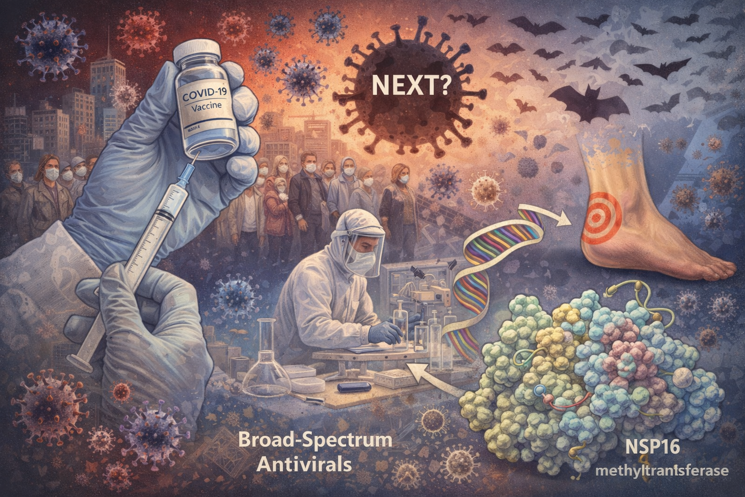
Authors: Ilham M. Alshiraihi, PhD; Gerald L. Klein, MD; Mark A. Brown
Abstract
With the approval and distribution of demonstrably safe COVID-19 vaccines bearing exceptionally high efficacy profiles, it may be tempting to envision a return to “normal” in the coming months. However, if there is one lesson to be learned from the ongoing pandemic, it is that, in a world of evolving zoonotic viruses, we must be better prepared for the next deadly outbreak. While the acute nature of the COVID-19 pandemic demanded a highly specific approach, it is advisable to consider the breadth of seemingly endless possibilities in our approach to managing the next inevitable occurrence of an outbreak. Though there is little chance of discovering a “magic pill” to combat all future pathogens, the highly conserved nature of non-surface viral proteins exposes an “Achilles’ heel” in the structural genome of viral pathogens. Herein, we consider the potential of targeting such proteins to develop broad-spectrum therapeutics for the future. To illustrate this point, we outline the therapeutic potential of targeting the nonstructural protein 16 methyltransferase, which is conserved across most coronaviruses.
Improving Clinical Trial Safety
There is a dire need to establish transparent medication safety guidelines and to assure the public of the industry’s commitment to this task. Clinical investigators and their staffs who conduct clinical trials often do not understand some of the regulatory methodology used to assess medication safety, particularly with regard to the assignment of causality to drug-related Serious Adverse Events. Yet, this is critical to establish the safety of medication. The medical monitor in a clinical trial can help educate and guide investigators in a clinical study with regard to accepted causation assignment. Since there is a paucity in the literature regarding medical monitoring of clinical trials, this article will help to fill this gap, and to provide a guide to establishing proper causality of an adverse event. This open access article can be found at Medical Monitoring of Clinical Research Studies.
Orthotopic grafting of decellularized human nipple: Setting the stage and putative mechanism of healing

A complex skin structure (such as a nipple) can be successfully decellularized under conditions that prevent extracellular matrix crosslinking or undue matrix degradation [1]. This treatment removes cellular antigens, thus mitigating immunorejection concerns and enabling allogeneic transplantation for nipple reconstruction after mastectomy. Non-human primate studies have shown that host-mediated re-vascularization and re-epithelization of the decellularized nipples occurs within six weeks and nipple projection is maintained over the same timeframe [1]. The mechanisms by which a decellularized graft located on the surface of the body heals are incompletely understood, but are likely to follow a similar path to decellularized allografts that are implanted within the body, with some modifications. The following is a description of probable temporal events leading to healing under this circumstance.
MedSurgPI is excited to tell the story of one of our innovative clients...

Keeping the Passion for Improving Patient Health Outcomes Moving Forward!
Esther M Alegria, Chief Executive Officer at APIE Therapeutics
My passion began in 1980 when, as a two-year college graduate, I joined the pharmaceutical industry in the island of Puerto Rico. The sense of contribution to my family, friends and every single user of the drug products we produced was embedded in every cell in my body. There has never been a question about what I should do in life.
This passion gave me the strength to go back to college and pursue a Ph.D. in Chemistry. Did I mention doing this while raising my two children, moving to a new country, and learning to speak English at the same time? Well that story is for another time. After completing my Ph.D. in Chemistry and an NIH Post-Doctoral assignment, I reentered the industry, in a Research and Development capacity. This path took me to Wyeth Vaccine R&D where I became closely involved in the development of Prevnar® and Meningetec® vaccines, from pre-clinical stage to commercialization. The journey, the learnings, and my mentors along the way were awesome and huge contributions to who I am today! I was honored to join each of my four grandchildren at the pediatrician’s office when they got their Prevnar® vaccination (I had to be there and witness it!).
My next big move was joining Biogen where my passion for moving forward new therapies to patients in need continued. In this journey I developed a second passion, seeing others develop their potential in the workforce. The satisfaction of managing and leading others on the right path that illuminates their skills and expertise has been a legacy that I treasure.
TODAY, I am not slowing down but starting the APIE Therapeutics, Inc journey. We have negotiated the exclusive worldwide intellectual property rights to RTI's (Research Triangle Institute International) decades worth of research on the Apelinergic System, Apelin/APJ receptor Agonists compounds portfolio, and pre-clinical studies of Apelin Agonist for the idiopathic pulmonary fibrosis (IPF) and heart failure (HF). APIE-Therapeutics Inc current strategy is to leverage the Apelinergic System Signaling Path to develop a portfolio of Anti-Fibrosis Therapies utilizing the RTI Apelin Agonists compounds portfolio (+800 compounds). The Apelin/APJ receptor is central to the pathophysiology of fibrosis, an underlying hallmark of many serious diseases. Drugs have been proposed for multiple diseases in pulmonary, cardiovascular and metabolic therapeutic areas. We have preclinical data in three specific therapeutic targets but intend to continue expanding the therapeutic targets within the Apelinergic System Signaling Path utilizing the RTI Apelin RTI Apelin Agonists compounds portfolio. The Idiopathic pulmonary fibrosis (IPF) indication is the pipeline program closest to being ready for human clinical trials.
We are excited to be moving this program forward given the strong need for better health outcomes; particularly, given the current pandemic and the observed increase in post-Covid-19 pulmonary fibrosis.
Unfortunately, there are only two available drugs in the market, and both provide limited health outcomes. The life span of IPF patients is 2-5 years. APIE-Therapeutics’ Apelin Agonists compounds novel mechanism of action for the IPF indication, has attracted the attention of top pulmonary/lung experts from Vanderbilt University Medical Center, University of Michigan Medical School and Weill Cornell Medicine, who have agreed to join our Scientific Board of Advisors. I am excited to have them be part of our team. We have a compelling preclinical data package and an experienced team of biopharma experts in pre-clinical and clinical development, regulatory, CMC and commercialization that I am confident will deliver on this program. There is a common motivation among all involved at APIE-T, a strong passion for bringing better health outcome to patients suffering from debilitating and fatal diseases.
We are launching an investor funding round this fall to move the IPF program into human trials. I am confident we will be conducting clinical trials next year!
Plakous Therapeutics extends research through agreement with Mayo Clinic

WINSTON-SALEM, N.C., August 25, 2020– Plakous Therapeutics, Inc. announced that it has signed a know-how license agreement with Mayo Clinic. The multi-year agreement will focus on understanding the results of a natural history study of the factors associated with necrotizing enterocolitis (NEC), a rare disease affecting premature babies. Plakous has received Orphan Drug and Rare Pediatric Disease designations from the Food and Drug Administration (FDA) for the prevention of NEC in premature babies born before 34 weeks of pregnancy. Plakous seeks to prevent NEC with Protego-PD™ by accelerating intestinal maturation of premature infants.
"Through this collaboration with Mayo Clinic, we will improve our understanding of NEC. As a leader in clinical outcomes research, Mayo Clinic is the ideal collaborator to seek the risk factors associated with this rare pediatric disease. We are excited to interface with leading neonatologist, William A. Carey, M.D., and leverage the breadth and depth of his experience with this research,” said Robert Boyce, Chief Executive Officer of Plakous Therapeutics.
“This study will provide valuable insights for the development and design of our clinical trials in NEC. This is a critical step in the advancement of our Investigational New Drug filing for Protego-PD™, an orally delivered acellular biotherapeutic developed by Plakous from postdelivery placentas,” said Boyce.
About Necrotizing Enterocolitis (NEC) NEC is a devastating disease caused by inflammation and lack of development of the intestine. More than 90% of the 6,000 annual cases in the United States occur in very low birth weight babies, babies born weighing less than three pounds. NEC carries a 30% mortality rate. Managing NEC consumes 20% of the $5 billion annual neonatal intensive care unit expenditures plus an estimated $4 billion in hospital costs for subsequent treatments.
About Plakous Therapeutics, Inc. Plakous Therapeutics is a biotherapeutic company dedicated to researching and developing placenta derived regenerative therapies to improve patient outcomes and reduce health care costs. For more information please visit the company’s website at plakoustherapeutics.com.
Orthotopic Grafting of Decellularized Human Nipple: Setting the Stage and Putative Mechanism of Healing

A complex skin structure (such as a nipple) can be successfully decellularized under conditions that prevent extracellular matrix crosslinking or undue matrix degradation (1). This treatment removes cellular antigens, thus mitigating immunorejection concerns and enabling allogeneic transplantation for nipple reconstruction after mastectomy. Non-human primate studies have shown that host-mediated re-vascularization and re-epithelization of the decellularized nipples occurs within six weeks and nipple projection is maintained over the same timeframe (1). The mechanisms by which a decellularized graft located on the surface of the body heals are incompletely understood, but are likely to follow a similar path to decellularized allografts that are implanted within the body, with some modifications. The following is a description of probable temporal events leading to healing under this circumstance.
Nipple Reconstruction
At some relevant timepoint after mastectomy and breast reconstruction, a woman will elect to have nipple reconstruction. After a review of all possible techniques, including tattooing, local flap rotation, autologous full thickness skin grafting from labia, and allogeneic or xenogeneic implants, it becomes clear that all are flawed in some way that prevents the accurate reproduction of a nipple’s 3D structure, pliancy, and color. A proposed new solution is to take advantage of the extensive evidence that a decellularized allogeneic or xenogeneic graft, an acellular extracellular matrix scaffold, can serve as the nidus for a healing event when implanted in a human (2). Although such scaffolds have long been derived from dermis, small intestinal submucosa, bladder and other organs, recent work has shown that deceased human donor nipples can be successfully decellularized and, when implanted on the surface of the body in a non-human primate model, exhibit complete healing within six weeks. Planned clinical studies will bring this technology to humans, where longed for accurate and complete nipple restoration can be evaluated.
When a woman elects nipple reconstruction with a decellularized human nipple, it is anticipated the following sequence of surgical events will occur. First, she will undergo presurgical evaluation to ensure that her reconstructed breast(s) have healed properly and bear no residual inflammation. Prior to surgery, she will be consulted regarding the relative size and placement of her new nipple(s). Markings will be made to denote these dimensions and positions and standard preparations for surgery will be made. Whether under local or general anesthesia, the breast skin will be prepped with an antiseptic, and prophylactic antibiotics will likely be administered. The surgeon will then remove the epithelial layer of the skin in the marked nipple location(s) on the breast(s), exposing the vascularized dermis, below. Punctate bleeding will likely require control by pressure or electrocautery to create a blood-free recipient field. The decellularized nipple(s) and surrounding areola will then be cut to the desired shape and sewn onto the breast(s) with either interrupted or continuous sutures. Wound dressings will be placed to encompass the nipple(s) but not compress the projecting nipple, itself. It is clear from the outset that this form of decellularized scaffold placement on the human body differs from other scaffold implantations in that it faces a vascularized interface only on one side. So, what is the likely pathway to its healing response?
Healing of Intact and Acellular Grafts
There will likely be some similarities to the healing responses of other living and non-living materials that are placed on a de-epithelialized skin base. Comparing this procedure to an autologous skin graft, it is expected that similar inflammatory steps would occur, but a salient difference is that the intact autologous graft contains micro vessels that would attach quickly to host tissue in a process known as inosculation, bringing nearly immediate life (within days) back to the skin that has been harvested (3). While not obviating interface inflammation and its effects (such as hypertrophic scarring in some cases), re-vascularization of autologous tissue such as a skin graft has the net effect of reducing the period of inflammation, since stress cytokines are no longer being sent from the tissue. In the case of an intact allogeneic or xenogeneic skin graft that is applied to de-epithelialized skin, which is common in burn treatment, inosculation will occur in a similar manner, leading to an apparent healing response that is soon truncated as an immune response develops and the graft is sloughed (4). Although this course of treatment does not lead to complete wound healing, the benefits of this application are twofold. First, the skin provides an essential moisture and infection barrier while the patient is stabilized for subsequent autografting. Second, the highly inflammatory immune response signals the underlying tissue to form a rich vascular network known as granulation tissue that serves as an optimal substrate for subsequent autografting (4).
There are many commercial products (Alloderm® from Allergan being one) that are composed of extracellular matrix proteins such as collagen and elastin (and in some cases fibroblasts and epithelium – Apligraf® from Organogenesis) that mimic the matrix component of skin grafts, whether autografts, allografts, or xenografts. By providing a milieu within which a mild immune response can operate, they often lead to the development of granulation tissue, even as they are eventually resorbed, leading to an improvement in subsequent autologous skin grafting. In addition, they have been used in nipple reconstruction to achieve some or all of the desirable features mentioned earlier, though with unpredictable and often disappointing results.
Effect of Decellularization on Healing
The entry into clinical care of the decellularized nipple allograft is therefore not without precedent, but some steps will be required to ensure that healing of such structures can proceed optimally. This begins with the preparation of the tissue itself. It has become clear in work with other decellularized tissues that the processes of decellularization (including in some cases, terminal sterilization) vary widely and can have substantial impact on the healing properties of matrices (5). The decellularization process must be harsh enough to remove cellular antigens, but mild enough to retain the native extracellular matrix structure, which is critical for providing the proper signaling and support structure to ingrowing cells. If decellularization is incomplete such that cellular antigens remain in immunogenic quantities, undesirable inflammation will be exacerbated, which can slow or prevent healing of the graft. But, if the extracellular matrix is damaged or crosslinked (an outcome that is particularly associated with gamma irradiation sterilization) by excessive chemical or mechanical preparation, the graft will also be less serviceable to the healing process.
Badylak points out that all scaffolds are eventually replaced by host tissue, but that depending upon modes of preparation, this can follow one of two pathways (5). When a scaffold is decellularized to retain its native structure, contains no cellular antigens and is not from an overly aged source, it undergoes a controlled, scar-free replacement process known as “Constructive Remodeling.” Conversely, if the decellularization is incomplete, too harsh, or terminal sterilization creates unnatural crosslinking, a highly inflammatory process is instituted, leading to scar tissue formation. This is known as “Deconstructive Remodeling.” Therefore, for a decellularized nipple allograft to have the potential to be replaced in situ and retain its 3D structure, it will be critical that substantial attention be paid to its preparation process after recovery.
Healing of an Externally Placed Acellular Nipple Graft
Now, assuming that such an optimal preparation has been achieved, what are the likely biological responses to a decellularized nipple allograft from the moment of attachment to a woman’s breast? Initially, despite the achievement of gross hemostasis at the interface during surgery, a thin veneer of fibrin is likely to be present beneath the graft. Platelets, attached to the fibrin through their GPIIB-IIIa receptors, will have been stimulated to release an array of cytokines that will recruit neutrophils and macrophages to the scene. As the latter cells enter the area and probe the underlying surface of the graft, they will begin work in three directions. First, they will sense the need to scavenge the fibrin and platelets and will release cathepsins and other lytic peptides. Second, the macrophages will predominantly be of the M1 or pro-inflammatory types and will begin releasing immunomodulating agents to recruit additional macrophages, neutrophils and eventually, fibroblasts. Third, the macrophages will also sense the low oxygen tension at the tissue interface and emit vasculogenic cytokines such as vascular endothelial growth factor (VEGF) and basic fibroblast growth factor (bFGF) (5).
As capillaries begin to proliferate at the undersurface of the graft, and as debris is cleared away, these vessels will begin to probe the graft itself, seeking to attach themselves to the graft by binding to preserved extracellular matrix anchors via integrins on their cellular membranes. Blood flow will eventually contribute endothelial progenitor cells to the site, leading to vascular ingrowth along the oxygen gradient that is present. Vascular pericytes that bear mesenchymal stem cell properties will then, in conjunction with macrophages, govern the early population of the graft and augment the recruitment of fibroblasts. In addition, for a breast mound that remains sensate after reconstruction, there is the potential for axons to enter the graft in parallel with the blood vessels. If the tissue preparation has elicited a very minor inflammatory response at this stage, a macrophage conversion will occur from the pro-inflammatory M1 state to the anti-inflammatory M2 state. The latter macrophages will then begin to release substances such as growth factors that elicit a Th2 (T helper cell) response, which “manages” the rate and spatial deposition of fibroblasts and also controls their production of extracellular matrix proteins, such as collagen, fibronectin, vitronectin and elastin, in amounts and orientations that respect the configuration of the decellularized matrix that they are slowly remodeling through replacement. This process will occur until an emerging vascularized border is present at the edge of the implant.
At this stage, epithelial cells at the border of the graft will have a toehold on the vascularized matrix and can begin to divide and migrate toward the center of the nipple as a function of the accretive vascularization of the implant. The migrating epithelial cells will likely include melanocytes when present. While fibroblast proliferation and volumetric remodeling of the graft itself continues to be governed by M2 macrophages and pericytes, the epithelium will continue to cover the nipple until this is complete. At that point, oxygen tension within the remodeled and now re-vascularized (and potentially re-innervated) nipple becomes normal and macrophages substantially recede, except for a population that will govern final remodeling of the nipple structure, an event that will be augmented by their mechanotransduction receptors as they are exposed to normal orthotopic breast stresses (6). When remodeling has achieved an “equal and opposite” relationship to those breast forces, remodeling will stop and potentially, some sensation will recover. Depending upon the degree of melanocyte participation in the epithelial migration (or, depending upon conversion of epithelial cells to melanocytes), the nipple will exhibit a hue that is reflective of their participation. In some cases, nipple tattooing may be performed to augment hue after remodeling has been complete. If re-innervation does occur, it is likely that the quality of sensation will continue to mature for some time after complete remodeling.
This constitutes a putative mechanism for the engraftment of a decellularized nipple allograft that is likely to follow the principles of engraftment of other foreign, decellularized tissues, albeit in the context of a unique placement on the surface of the human body.
References
1. Graham, D, et al, Abstract P6-14-13: New Approach To Nipple Reconstruction: In Vivo Evaluation Of Acellular Nipple-Areolar Complex Grafts, Cancer Res February 15 2020 (80) (4 Supplement) P6-14-13; DOI:10.1158/1538-7445.SABCS19-P6-14-1
2. Badylak, SF, The Extracellular Matrix as a Biologic Scaffold Material, Biomaterials, 28:25, 3587-3593, 2007.
3. Laschke, MW, et al, Inosculation: connecting the life-sustaining pipelines, Tissue Engineering, Part B: Reviews, 15:4, 455-465, 2009.
4. Yamamoto, T, Skin xenotransplantation: Historical review and clinical potential, Burns, 44:7, 1738-1749, 2018.
5. Badylak, SF,Decellularized Allogeneic and Xenogeneic Tissue as a Bioscaffold for Regenerative Medicine: Factors that Influence the Host Response, Annals of Biomedical Engineering, 42:7, 1517-1527, 2014.
6. Chamberlain, MD, et al, In Vivo Remodelling of Vascularizing Engineered Tissues, Annals of Biomedical Engineering, 43 1189–1200, 2015.
Current Therapeutic Research Special Edition on Medication Adherence

A special edition of Current Therapeutic Research, an Elsevier journal, will be dedicated to Medication Adherence. We have the pleasure of being guest editors of this special edition. If you are interested in being an author of an article for this edition there is an opportunity to rapidly communicate the results of your research, or thoughts, to a diverse, international readership of scientists and clinicians. Current Therapeutic Research is a well-regarded, peer-reviewed, rapid publication Elsevier journal in the field of clinical trials and other developments in drug therapy. CTR has significant reach, with over 15,000 downloads and page views per month via Elsevier’s ScienceDirect and journals platform. Once published, all content will be immediately available and freely accessible online.
If you are interested, I would ask you to submit an original article, review, or commentary. This manuscript should be submitted through our electronic submission system (Editorial Manager). It should adhere to the format guidelines in the Guide for Authors. When you are ready to contribute a manuscript, the Editorial team of Current Therapeutic Research will work with you and provide assistance as you prepare and submit your manuscript. Please don't hesitate to communicate directly with me, gklein@medsurgpi.com , the Managing Editor (Judy Pachella; J.Pachella@elsevier.com); or the Editor-in-Chief (Dr. Phil Walson; pwalson1@aol.com) if you have any questions.
THE COMPOSITE VIRTUAL MEDICAL OFFICER

There is a global shortage of physicians and experienced biopharmaceutical and medtech physicians. [1] High salaries as well as other costs associated with these positions are expensive for small companies and stretch the budget in larger corporations. A physician’s medical expertise may be limited by their training, knowledge, experience, and abilities. An effective company neurologist may not be able to help if you have just licensed a dermatologic product. Many doctors may excel at product development, but not be that astute in medical affairs and assisting the commercial organization. Conversely, they may be very effective in medical affairs, but not as knowledgeable in medical monitoring and pharmacovigilance.
This need has created the Composite Virtual Medical Officer (CVMO), offered exclusively at MedSurgPI. A full time CVMO consists of two or three experienced pharmaceutical physicians, who provide a comprehensive package of services, talents and abilities. One MD serves as the project managing physician, ensuring the additional doctors are kept well-informed of all the activities. They serve as one cohesive unit, working together on your project to provide a comprehensive product, often superior to the abilities of one person. This composite virtual medical officer can be full or part time and there are no interruptions to activities with vacation or sick days. The CVMO provides the luxury of having two or three physicians on your team for the price of one.
Advantages of a CVMO:
Greater depth of knowledge and experience than a single hire
Multiple skills in functionable capacity to be an expert in development, monitoring, medical affairs, interacting with the FDA, and pharmaceutical business executive
Full or part time
Economical
Total coverage for vacation or sick leave
Virtual or in-office meetings
Able to be in two places at once (when necessary)
No overhead costs for office, equipment or paid benefits
Expertise in diverse functions and therapeutic specialties
Greater flexibility with multiple options for the following:
+Permanent position
+Immediate interim function while searching for a full-time medical officer
+Changing your lead CVMO as the functional role changes
[1] Degen et al. Human Resources for Health (2015) 13:74
MAINTAINING PRODUCT DEVELOPMENT MOMENTUM IN A CHALLENGING TIME
It is important that biotech and device companies continue their critical research and development during these chaotic times. Many small companies are having difficulty raising funds and may be getting short of cash. Consulting companies have the flexibility to work remotely with these companies and can consider delayed payments, use of equity, or other innovative means to compensate them for their assistance while keeping momentum strong and morale high. MedSurgPI, LLC fulfills this role by providing virtual medical support for product development, medical monitoring, medical affairs, and serving as Chief Medical Officers. Should you require these services, please contact us at the following:
Drugs, Devices and Biologicals: gklein@medsurgpi.com
Medical Monitoring and DSMB activity: rmorgan@medsurgpi.com
Find us at: www.medsurgpi.com
MedSurgPI, LLC provides remote Medical Officer and Commercial Chief Medical Officer Services during this critical time of the Covid-19 Outbreak.
Social distancing during the Covid-19 crisis is adversely affecting the ability of companies to continue biomedical product development and to plan and execute clinical trials and medical affairs activities. While this crisis will eventually abate (albeit with an uncertain timeline) virtually located professionals are needed in the interim to continue to support these critical development activities. Research Triangle Park, NC-based MedsurgPI, LLC offers 20+ experienced Physician Associates from all areas of Medicine to provide comprehensive remote medical officer and medical monitoring services to companies worldwide. These services also include strategic product development oversight, clinical trial planning, Contract Research Organization sourcing and management, Medical Communications, interim management, support for fundraising, Key Opinion Leader recruitment and facilitation and medically relevant corporate representation. Should your organization require these skills on a remote basis in the near future, please contact MedsurgPI, LLC and we will respond immediately to support you.
Gerald L. Klein, MD, Principal MedSurgPI
www.medsurgpi.com
gklein@medsurgpi.com
Australian R&D Tax Incentive
How The Australian Research and Development (R&D) Tax Incentive Works
Peter C. Johnson, MD and Gerald L. Klein, MD
Principals, MedSurgPI, LLC
Introduction
In order to foster greater corporate R&D investment to spur jobs and innovation, the Australian government adopted the R&D Tax Incentive system in 2011. While its stipulations have gradually changed over the years, it remains a generous incentive for companies large and small to invest in R&D in Australia. The plan has two broad elements, one aimed at supporting companies having less than 20 million in annual revenue (these are Australian dollars, equivalent to 13.4 million US dollars) and those having greater than 20 million in revenue. The incentive applies not only to native Australian companies but also to foreign companies that establish a bona fide Australian presence and comply with Australian business guidelines as set by the Australian Securities and Investments Commission (ASIC: https://www.investopedia.com/terms/a/australian-securities-and-investments-commission-asic.asp). A straightforward set of FAQs regarding these requirements can be seen here: http://cosec.com.au/frequently-asked-questions/faqs-on-australian-companies/. Additionally, types of R&D activities that are eligible for the benefit are carefully defined in order to prevent abuse of the system. These and current applications of the Incentive are outlined below:
R&D Expenditure Threshold
For all companies, the threshold R&D spending amount to which the Incentive applies is now 150M Australian dollars (100.5 million US dollars).
Companies Having Less Than 20 Million in Revenue
Such companies are entitled to a refundable tax credit equal to their corporate tax rate percentage plus 13.5 basis points. Given that the present corporate tax rate is 27.5%, their refundable tax credit as of the 2019 legislation is 41% of their R&D spend. The word “refundable” requires some explanation. For companies whose revenues impart no tax burden, they are entitled to a cash rebate of 41% of their R&D spend. If they have a tax burden, the 41% will initially be used to offset that amount of tax burden up to and including 41% of their R&D spend. If their tax burden does not achieve this level, the refundable incentive will first be used to offset their tax burden and the remainder distributed to the company as a cash rebate.
The Incentive also distinguishes between expenditures for clinical trials and for R&D that is not specifically a clinical trial expenditure (such as preclinical studies). There is a 4 million Australian dollar (2.68 million US dollar) cap on the latter activities but no limit on the Incentive as applied to clinical trial activities up to the R&D Expenditure Threshold of 150 million Australian dollars.
Some examples are shown in as follows, applicable only to companies having less than 20 million Australian dollars in revenue.
Example 1: Acme Medical spent zero dollars on Clinical R&D and six million dollars on Non-Clinical R&D. Its tax burden was zero so its tax offset is zero and it receives a cash rebate of four million dollars.*
Example 2: RNA Science spent one hundred million dollars on Clinical R&D and zero dollars on Non-Clinical R&D. Its tax burden was zero so its tax offset is zero and it receives a cash rebate of forty one million dollars.
Example 3: Mitochondrial Solutions spent zero dollars on Clinical R&D and ten million dollars on Non-Clinical R&D. Its tax burden was four million so its tax offset is four million and it receives a cash rebate of zero dollars.*
Example 4: Atlas Diagnostics spent fifty million dollars on Clinical R&D and six million dollars on Non-Clinical R&D. Its tax burden was two million so its tax offset is two million and it receives a cash rebate of 19.7 million dollars.**
*Only the first 4M of any R&D tax offset for non-clinical trial related activities can be applied each year. Any remainder must be carried forward as a non-refundable tax offset (see below) that can be applied in future years. This limit does not apply to clinical trial activities.
*In this instance, non-clinical R&D spend refund, capped at 4M is partially applied (2M) to offset the 2M tax burden, leaving 50-2=48*.41=19.7M available as a cash rebate.
Companies Having Greater Than 20 Million in Revenue
Circumstances of application of the Incentive are quite different for larger companies. Companies having greater than 20 million in revenue are entitled to a non-refundable tax incentive equal to their corporate tax rate PLUS a sliding scale premium based on the level of intensity of their R&D expenditure. “Non-refundable” in this instance means that the Incentive rebate only applies to offsetting the company’s tax burden with no cash rebate. However, if the calculated tax incentive amount exceeds that of its annual tax burden, the residual amount can be carried forward as an applicable tax credit for future years.
The sliding scale R&D “intensity premium” (that is, above and beyond their corporate tax rate) is calculated as a function of the percent of a company’s total expenses that are represented by R&D expenses. For an R&D Intensity Range (that is, R&D expense as a percentage of total expenses) that is less than or equal to 4, the Incentive Premium (% of Total Expense) is 4.5. When the R&D Intensity Range is greater than 4 but less than or equal to nine, the Incentive Premium rises to 8.5%. Finally, when the R&D Intensity Range is greater than nine, the Incentive Premium rises to 12.5%.
The following provides some examples of how the program applies to entities having greater than 20 million in annual revenue. Note that corporations having greater than 50 million in annual revenue are subjected to a 30% corporate tax rate.
Example 1: Claro Science’s R&D expense is 1 million versus total expense of 40 million. It has 45 million in revenue. Its applicable corporate tax rate is 27.5%. Its tax burden is 12.4 million and its R&D expense as a percent of total expense is 2.5. Its Incentive Premium (% of total expenses) is 4.5. Its earned incentive tax offset is 18 million. Therefore, this year its incentive tax offset is 12.4 million and it is able to carry forward 5.6 million in incentive tax offset for future years.
Example 2: Adams Medical’s R&D expense is 3 million versus total expense of 50 million. It has 60 million in revenue. Its applicable corporate tax rate is 30%. Its tax burden is 18 million and its R&D expense as a percent of total expense is 6. Its Incentive Premium (% of total expenses) is calculated as 4.5(4)+8.5(2). Its earned incentive tax offset is 35 million. Therefore, this year its incentive tax offset is 18 million and it is able to carry forward 17 million in incentive tax offset for future years.
Example 3: Hong Kong System’s R&D expense is 9 million versus total expense of 135 million. It has 150 million in revenue. Its applicable corporate tax rate is 30%. Its tax burden is 45 million and its R&D expense as a percent of total expense is 6.7. Its Incentive Premium (% of total expenses) is calculated as 4.5(4)+8.5(2.7). Its earned incentive tax offset is 40.3 million. Therefore, this year its incentive tax offset is 40.3 million and it is able to carry forward zero dollars in incentive tax offset for future years.
Example 4: Pacific Bio’s R&D expense is 50 million versus total expense of 450 million. It has 500 million in revenue. Its applicable corporate tax rate is 30%. Its tax burden is 150 million and its R&D expense as a percent of total expense is 11.1. Its Incentive Premium (% of total expenses) is calculated as 4.5(4)+8.5(5)+12.5(2.1). Its earned incentive tax offset is 86.8 million. Therefore, this year its incentive tax offset is 86.8 million and it is able to carry forward zero dollars in incentive tax offset for future years.
*R&D Expenditure Threshold capped at 150M. See above. Also, the scale applies incrementally – that is R&D expenditure up to 4% is rewarded by a 4.5% inventive premium, that portion of R&D expenditure between 4% and up to 9% is rewarded by an 8.5% incentive premium and that portion of R&D expenditures > 9% of total expenses is rewarded by a 12.5% incentive premium.
In Country Guidance
Support for companies seeking these benefits – especially foreign entities with an Australian presence – can be assisted through the complexities of compliance and application by local consultancies having expertise in these areas. A Google search entitled “Australian firms providing R&D tax incentive support” provides a starting point for the identification of such groups: https://search.yahoo.com/search?fr=mcafee&type=E211US667G0&p=Australian+firms+providing+R%26D+tax+incentive+support. Australian Contract Research Organizations are also adept at guiding clients to such support organizations.
Overall, the Incentive is jointly administered by Innovation and Science Australia (ISA) and the Australian Taxation Office (ATO), the former responsible for registering R&D activities while the latter manages the rules regarding who is eligible and overseeing acceptable costs. Background information and guidance regarding eligibility and application to the program can be found here: https://www.business.gov.au/grants-and-programs/research-and-development-tax-incentive
The revised legislation governing the program as introduced into the Australian Parliament in 2019 can be found here: Treasury Laws Amendment (Research and Development Tax Incentive) Bill 2019.
MedSurgPI Partnership Announcement

MEDSURGPI PARTNERSHIP ANNOUNCEMENT WITH DEVICIA FOR EU DEVICE EXPERTISE
MedSurgPI is expanding our services through our unique partnerships with experienced, dedicated, entrepreneurial consulting companies. In light of the current significant regulatory changes in the EU, increasing our regulatory and clinical expertise in Europe is essential. For that reason, we are proud to announce our new relationship with Sweden-based Devicia, a full-service medical device exclusive CRO. As members of Technical Committees and nominated as experts in international working groups writing medical device standards (such as ISO 14155), we will be able to provide our clients with an industry perspective and first-hand expert advice.
Through this collaboration, MedSurgPI will provide:
· MDR & IVDR transit and compliance
Devicia has extensive knowledge of regulatory changes currently taking place in the EU with the implementation of medical device regulation (MDR) and in-vitro diagnostic device regulation (IVDR). Through the collaboration, MedSurgPI and Devicia can assist with regulatory strategy & gap analysis, technical files and design dossiers, classifications, regulatory submissions etc.
· Clinical Evaluations
A key component in getting or maintaining a CE mark is the Clinical Evaluation Report. All medical devices on the EU market must perform a Clinical Evaluation according to MEDDEV 2.7/1 Rev 4. Devicia’s experienced medical writing team performs clinical evaluations according to the current guidelines and fulfils the requirements of the role of a clinical evaluator.
· EU Device Clinical Investigation
Devicia provides full-service support for all kinds of clinical investigations (single center, multicenter multinational RCT’s, registries etc.) both before and after CE mark. Devicia also supports clients with the new MDR requirements for Post Market Clinical Follow up (PMCF). Devicia is nominated as a Swedish expert in the ISO/Technical Committee 194 Working Group 4 governing clinical investigations of medical devices in humans.
· Licenses for ISO 14155 Clinical QMS
ISO 14155:2011 is specifically tailored to the requirements for medical device clinical investigations. Through the collaboration, MedSurgPI can offer access through licenses to SOP’s, templates and forms to be able to conduct a clinical investigation according to ISO 14155:2011.
gklein@medsurgpi.com / 919-930-9180